2026 HOPE LIVES GALLERY
2026 marks the 5th annual Hope Lives: Art for ALS exhibition at the San Francisco Women Artists Gallery. Melissa Stephens began curating this show in 2022 to honor her sister, Lynn Giovannelli, who was diagnosed with ALS in July 2020. Lynn passed away in 2023, but Stephens continues to educate and fundraise for ALS research. She hopes that someday soon her efforts
will help find a cure for people with ALS.
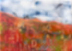
This year's exhibition is: Colors from Grief to Light - a collection of Melissa Stephens' encaustic paintings that explore feelings of loss, highlight memory and dive into her ongoing
transition from mourning a death to celebrating a life.
Exhibition Dates: May 5 - 29, 2026
Opening Reception: Saturday, May 9th from 2 - 4 pm
In addition to the exhibition, Melissa Stephens is hosting a Gelli Plate Printing Workshop on Saturday, May 16th from 10 am - Noon. This family friendly class will have limited seating, so register early!